UChicago Experiment Guides
Documentation to help you try new thingsSolvent Suppression 1: Single Peak at Center of Spectrum
Preferred technique = “Excitation Sculpting”
For full method, see p. 29 of Bruker’s “Guide Book – Advanced NMR Experiments” manual.
Outline:
- Acquire a normal 1D 1H spectrum without suppression. It’s convenient to use NS=1 to reduce experiment time. It’s important to automatically set the gain.
- Don’t expect to see peaks close to the solvent peak
- Do expect automatically set RG between 1.0 and 5.0
- Set the transmitter on the solvent peak
- Zoom in on the solvent peak at high-resolution. Make it occupy most of your window.
- Click the “set transmitter frequency” tool – the one that looks like a little lightning bolt.
- Using the red cursor that appears, set it precisely at the center of your solvent peak.
- Click it when the red line is properly positioned
- You’ll see a window pop up with a new value for O1
- To be safe, write down the value for O1, in Hz.
- Click OK to set the O1 value.
- Acquire another 1D 1H spectrum with the new O1 value. Click “Go” and overwrite the old spectrum.
- The resulting spectrum should be shifted so the solvent is in the center.
- If desired, adjust SW to include all the peaks in your spectrum; it’s possible you need to make SW larger than default to include them all.
- Type “iexpno” to create a new experiment with the same parameters as your original.
- Try a new SW, acquire a spectrum, check out the resulting spectrum.
- Settle on a final SW
- Set up a preliminary solvent-suppressed experiment
- Type “iexpno” to copy the existing good parameters (with desired O1 and SW) to a new experiment.
- set PULPROG = ZGESGP
- set P12 = 2000 microsec
- set GPNAM1 = SMSQ10.100, set GPNAM2 the same
- set SPNAM1 = Squ100.1000
- Pull down the “Prosol” tab to “pulsecal” to perform a single-scan pulse calibration
- Type RGA to automatically set the receiver gain. You should expect it to be 512 to 2050.
Bruker Topspin Manuals
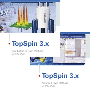
Bruker’s Topspin 3.X manuals offer highly useful step-by-step guides to setting up experiments. If you want to try something new, please feel free to explore these manuals and follow the instructions.
Basic: Introduction to 1D and 2D NMR Methods
Next Step: Advanced NMR Methods
If you have any questions or would like additional orientation, please contact the facility manager.

Experiments Available in Automation
(400-1 & new 400-2)
Experiment times do not include overhead for sample changing, tuning, locking, shimming, or setting gain.
Parameter Set |
Default Time |
Description |
Pulse Sequence |
| PROTON1 | 20 sec | 1D 1H, single scan with 90° excitation, 17 sec delay to allow for full relaxation. | zg |
| PROTON8 | 36 sec | 1D 1H, 8 scans, optimized for maximum signal and reliable integrals. For all multi-scan 1H 1D spectra with more scans, use these parameters and change NS. See blog post for optimization details |
zg30 |
| CARBON | 10 min | 1D 13C, 128 scans. Provides OK S/N for 10 mg samples. Change NS, but not D1, AQ, or P1, for increased signal. See blog post for optimization details |
zgdc30 |
| CARBON_3.5hr | 3.5 hr | Same as CARBON, but with NS=4096. Night queue only |
zgdc30 |
| DEPT135 | 2 min | 1D 13C DEPT135. 13C-detected, but signal enhanced via 1H magnetization. Only 13C atoms with 1H’s attached are observed. When properly phased, CH and CH3 13C’s are positive, CH2’s are negative. | deptsp135 |
| COSY-opt | 8.5 min | 2D COSY (COrrelation SpetroscopY), Indirect TD fixed at 200 increments. | cosygpppqf |
| HSQC-opt | 10 min | 2D 1H-13C HSQC (Heteronuclear Single Quantum Coherence), indirect TD fixed at 128 increments; multiplicity-edited so CH and CH3 peaks are positive and CH2 are negative (as in a DEPT135 spectrum), sensitivity improved over standard. Gives very good signal levels on samples less than 10 mg. | hsqcedetgpsisp2.4 |
| HMBC-opt | 22 min | 2D 1H-13C HMBC (Heteronuclear Multiple Bond Correlation), indirect TD fixed at 256 increments, filters eliminate one-bond correlations. Gives good signal on 10 mg samples. | hmbcetgpl3nd |
| NOESY-opt | 2.25 hr | 2D NOESY (Nuclear Overhauser Effect SpectroscopY) for detecting/measuring 1H-1H through-space contacts and chemical exchange, indirect TD fixed at 256 increments, default mixing time D9 = 0.6 sec (but can be modified). Sophisticated pulse sequence elements reduce DQCOSY-like +/- zero-quantum coherence (ZQC) artifacts. With positive diagonal peaks, negative crosspeaks indicate NOE and/or chemical exchange. Gives good data on 10 mg samples in the time specified. Night queue only. |
noesygpphzs |
| ROESY-opt | 52 min | 2D ROESY (ROtating frame noESY) for detecting/measuring 1H-1H through-space contacts and chemical exchange leveraging rotating-frame magnetization, indirect TD fixed at 128 increments, default mixing time is 0.6 sec. Also known as the “EASY-ROESY”, this “jump-symmetrized” ROESY reduces TOCSY artifacts and improves sensitivity. With positive diagonal peaks, positive crosspeaks indicate NOE contacts, and negative crosspeaks indicate chemical exchange. Gives good data on 10 mg samples in the time specified. Night queue only. |
roesyadjsph |
| TOCSY-opt | 42 min | 2D 1H-1H TOtal COrrelation SpectroscopY with DIPSI spin lock for detecting peaks in the same network of 1H-1H coupled spins, indirect TD fixed at 128 increments, default mixing time D9 = 0.08 sec that can be changed by user. | dipsi2gpphzs |
| HSQC-TOCSY-opt | 15 min | 2D 1H-13C correlation combined with TOCSY; with 1H direct detection axis, 13C indirect detection. Each 13C resonance is coupled to the 1H covalently attached AND the 1H resonances in its 1H-1H spin-coupled network; excellent for assigning 1H peaks overlapped in 1H, COSY, and TOCSY spectra. Indirect TD fixed at 200 increments, default mixing time D9 = 0.045 sec. | hsqcdietgpsisp.2 |
| CHARACTERIZATION | 55 min | Sets up PROTON8, CARBON, DEPT135,COSY,HSQC,HMBC in one click | various |
| FLUORINE | 3 min | 1D 19F with 90° excitation and 1H decoupling during acquisition. Uses a special spin-echo pulse sequence to eliminate background 19F signals from Teflon. See blog post for optimization details. |
zgfhigqn.2_se.jk |
| FLUORINE90 | 3 min | 1D 19F with 90° excitation and no 1H decoupling or background-reducing pulse sequence. | zg |
| PHOSPHORUS30 | 1 min | 1D 31P with 30° excitation and no 1H decoupling | zg30 |
| PHOSPHORUS30_1Hdec | 1 min | 1D 31P with 30° excitation and 1H decoupling. | zgig30 |
| std11B.SMP | 1 sec* | 1D 11B with 90° excitation and no 1H decoupling, *NS=1 scan by default | zg |
| LITHIUM7_3min | 3 min | 1D 7Li with 30° excitation and no 1H decoupling | zg |
| HN-HSQC-opt | 11 min | 2D 1H-15N HSQC with indirect TD = 200 increments. Gives OK spectra on 10 mg samples | |
| HN-HMBC | 40 min | 2D 1H-15N HMBC with indirect TD = 256 increments. Gives good spectra on 10 mg samples. Night queue only. |
|
| PROTON_T1 | 2 hr | Pseudo-2D T1 measurement experiment with 16 time points. For processing instructions, click here to access Bruker’s Advanced 1D and 2D Experiments guide. Night queue only. |
t1ir |
x
